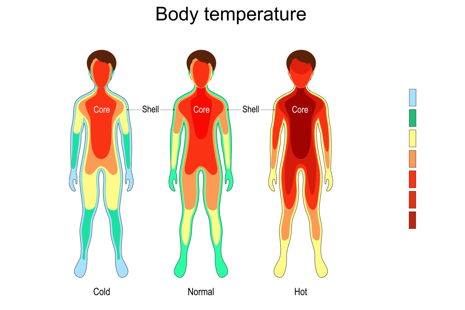
A medical imaging technique that uses infrared to capture images.

Why Choose Thermography?
Thermography is a sophisticated diagnostic modality that examines skin temperature changes on the surface of the body. It is a non-invasive, radiation-free approach that analyzes the skin – the largest, most intelligent, and easily accessible organ of the human body. The technique uses an infrared camera to detect and record the temperature variations that occur on the skin surface, which can provide valuable information about underlying physiological functions and pathological conditions. Thermography is a highly sensitive tool that can be used to monitor a wide range of health concerns, including breast cancer, vascular disease, neurological disorders, and pain management.
Our skin is like the central processing unit of our body. It functions as a central communication hub that seamlessly connects and shares information between all bodily systems, including the immune, neurological, and endocrine functions and pathways. The skin contains valuable information, and thermography acts as a monitor by measuring and evaluating metabolic signals, telling the story as it happens.
Skin Communication
The integumentary system is a crucial organ that provides non-verbal cues about an individual’s physical well-being. These indications are visible to the naked eye and can provide insights into the individual’s overall health status. Color changes within the skin can indicate a range of conditions, including a lack of oxygen, exposure to toxic chemicals or radiation, traumatic injury, extreme temperatures, emotional stress, and embarrassment. Additionally, the skin can display signs of alcohol and tobacco abuse and various skin disorders, which can be symptomatic of underlying morbidity or psychological disorders.
While the naked eye may provide insight, more sophisticated diagnostic methods are available to analyze the state of the skin. Thermography can provide a more in-depth examination of the skin, revealing underlying conditions that may be invisible to the naked eye. This technology can be instrumental in identifying underlying health issues before they become symptomatic, enabling earlier intervention and treatment.
The skin undergoes a constant change that generates subtle voltage variations and thermography signals. These signals provide a unique and non-verbal form of communication that can be used to monitor the body’s health.
The human body possesses a remarkable capacity to self-maintain and self-heal, provided that it is not subjected to abuse. Infrared imaging, commonly known as thermography, offers a heightened sensitivity that enables the observer to monitor the body as it prioritizes problem areas. This technology is instrumental in inquiring about the body’s health status and examining the responses it produces. With the valuable insights gained, healthcare professionals can then proceed to investigate further with a more targeted approach.
Our skin is an intelligent, dynamic, and familiar organ that reveals a lot about our general health. Sometimes, without realizing it, we subconsciously evaluate people’s health by looking at their skin. Now, we have the ability to observe the skin for this purpose intentionally. Therefore, thermography is not just a device that detects heat but also an exciting tool for observing our skin.
Understanding How Thermograms Are Different
Thermography is an imaging technique that captures the physiological activity of the breast instead of just the structure. For instance, an X-ray of the heart will reveal its size and location within the chest, while an EKG will display how the heart functions. Similarly, thermography captures the functional activity of the breasts and provides an insight into their overall health.
Many tests can only detect an existing mass without being able to differentiate between a benign lump and a cancerous tumor. However, the formation of blood vessels needed to feed a tumor occurs many years before the tumor itself becomes visible. This vascular formation produces heat that can be detected using thermal imaging. If thermal imaging indicates the presence of heat, it could be the result of your body preparing to produce a cancerous tumor or that the existing mass is most likely cancerous.
It’s important to understand that there is a significant difference between healthy breasts and breast cancer. Knowing where you land on this spectrum can help maintain good breast health. Several factors can contribute to unhealthy breasts, such as dense and fibrocystic breasts, inflammation, calcifications, lymph congestion, thyroid dysfunction, hormone imbalances, and more. Ignoring these factors can lead to an unfavorable diagnosis in the future.
It is important to take steps to prevent diseases. One of the first things to do is to identify your risk factors. Once you know your risk factors, you can create a plan to improve your health and even reverse some existing health problems. Remember, knowledge is power, so take action towards a healthy and happy life!
Explanation of Thermography
This highly specialized medical imaging technique uses infrared to capture images of the body based on heat and blood vessel patterns. It can detect inflammation, infections, and other functional concerns throughout the body, including the breasts. While other breast imaging technologies like mammography, MRI, and ultrasounds are mainly used for screening and detecting breast cancer, thermography is a preventative tool that can help evaluate breast health.
Unlike other imaging technologies, thermography evaluates the physiology of the breast, not the anatomy. It can detect physiological changes in the breast as early as five to ten years before a tumor develops, making it an effective tool for early detection. By detecting changes early, simple lifestyle changes, diet, and supplements can be used to reverse these patterns quickly and prevent progression to cancer.
After a tumor has developed, thermography can show different patterns. Sometimes, the patterns can appear very worrisome, with high heat and an aggressive look. Other times, the patterns can seem harmless and even show a lack of heat if the tumor has suffered central necrosis.

Thermogram vs Mammogram
It is important to note that no single test is perfect for evaluating breast health. While useful, Mammography, MRI, US, and thermography should not be used in isolation. For decades, mammography was considered the “gold standard” and used as a stand-alone test. However, long-term studies have revealed that mammography’s effectiveness has been overestimated while its limitations have been underestimated. For instance, mammography can miss up to 50% of tumors, especially in women with high breast density. Additionally, it often identifies “suspicious” areas that require a biopsy, resulting in unnecessary biopsies 80% of the time. Similarly, the other tests are not foolproof and can also miss tumors.
Research studies have established the importance of conducting multiple tests to achieve the optimal level of sensitivity and specificity. In light of this, it is recommended that, in addition to thermography, an anatomical test, such as an ultrasound, be conducted annually, along with a physical examination. In the event of any symptoms, such as lumps, further investigation is crucial and should include a biopsy for a tissue diagnosis.
Other Services

IV Therapy

Aesthetics


